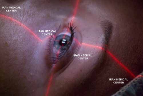
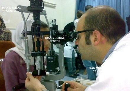
Ophthalmology is considered very important in Iran. Now all stages of medical and surgical ophthalmology are performed by Iranian experts in Iranian ophthalmology centers and hospitals. Ophthalmology services in Iran are higher than international standards. Iran ophthalmology centers use experienced specialists as well as advanced diagnostic and surgical facilities and technology to provide a wide range of services related to the visual system problems. In the case of visual disturbances associated with other systemic problems like diabetes, autoimmune and thyroid disease; the cooperation and teamwork of Iranian specialists can meet all the needs of the patients and provide care and treatment. Iranian hospitals offer the following ophthalmology services:
Cataract
Glaucoma
Macular degeneration
Corneal surface diseases
Eyelid and orbital diseases
Retinal diseases
Visual sensory impairments
Refractive defects
Strabismus
Permanent change of eye color
This is another application of artificial iris for cosmetic purposes. Artificial iris is available in different colors, and the surgeon implants the color desired by the patient. This color change is permanent. However, if for whatever reason, there is a need, the artificial iris can easily be removed.
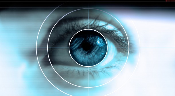
Everybody’s eye color is induced by color of the iris. Iris is the pigmented part of the eye, and its color depends on genetic attributes. Iris is normally, brown, black, blue, gray, or green, and generally depends on two pigments: melanin and lipofuscin (for some shades of green). In absence of any pigment (albinism), eye turns to the color of blood circulating in it.
Iris colors in different parts of the world are highly diverse, and in some people, it is made up of combination of two or more different colors. Most common eye or iris colors are: brown, black and chestnut, which comprise ¾ of people in the world. In Iran, most eyes are dark brown. Iris has an important role in controlling amount of light entering the eye, and functions to protect eyes against harmful sun rays. Some people have no iris or defective iris because of trauma to the eye, or diseases.
Artificial iris
Artificial iris is a fine, flexible non-toxic prosthesis that is compatible with body tissue. Artificial iris is made of similar materials used in manufacture of intraocular lenses. These lenses have been inside patients’ eyes for many years (for example: after cataract surgery). Therefore, they are made of harmless materials with no complications.
Corneal Collagen Cross-Linking with Riboflavin - UVX
Corneal collagen crosslinking (CXL) was developed in 1998 by Theo Seiler, MD, and has been shown in numerous clinical trials to strengthen the eye's clear surface (cornea) through the application of riboflavin, a form of vitamin-B2, followed by treatment with ultraviolet A (UV-A) light.
The two basic types of corneal crosslinking are:
Epithelium off, which means the thin layer covering the eye's surface is removed, allowing for faster penetration with liquid riboflavin. Transepithelial corneal crosslinking (epithelium on) is where the corneal epithelial surface is left intact, which requires a longer riboflavin loading time.
Cross linking with riboflavin and UV-A light has proven to be a first-line treatment for people with eye conditions such as keratoconus, pellucid marginal degeneration and corneal weakness (ectasia) after LASIK.
Cataract Surgery
In cataract surgery, the cloudy natural lens must be removed from the eye. After that, in most cases a permanent intraocular lens (IOL) implant replaces the natural lens to restore focusing power.
When to have cataract surgery often is a subjective decision, based on how well you are able to see during routine activities. You might be able to drive, watch television and work at a computer for quite a few years, even after you are first diagnosed with cataracts.
However, if you have cataracts, you may eventually start to notice "ghost" images and declining visual clarity, which can't be corrected withglasses or contacts. Colors may begin to look faded, too. If your functional vision is impaired significantly and it becomes difficult for you to perform your normal daily activities, it may be time for cataract surgery.
LASIK / Femto-LASIK
LASIK,the most frequently performed, the most accurate and the safest refractive eye surgery, is a two-step procedure that is performed on the cornea. The first step is cuttinga layer (a thin flap) of the corneal tissue. Not many years ago, in the classic LASIK procedure, this corneal lamella (flap) was prepared with a microkeratome (a surgical precision instrument with an oscillating blade), however nowadays the femtosecond laser prepares the flap–without any use of mechanical blades. This increases the accuracy and safety of the surgery to a large extent. Femto-LASIK is the combination of Excimer laser and Femtosecond laser, and is one of the best methods for correcting refractive errors.
Femto-LASIK is one of the newest methods of correcting myopia, hyperopia and astigmatism. Femtosecond laser is widely applied in ophthalmology, which includes all kinds of cutting the cornea and lenses, treatment of glaucoma and retinal deficiencies. Currently, it is mostly used in cutting the cornea during LASIK and it can be considered the superior LASIKmethod. Femtosecond laser is a megatrendcutting technology. Each Infrared femtosecond laser pulse is a few seconds (15-10 seconds), thus it causes no damage to the peripheral areas.
Femtosecond laser devices are equipped with soft wares by which the number, size, amount of energy and frequency of the radiatedpulses can be adjusted. This software can also determine the exact location of the radiation according to the required breadth and depth. So while using Femtosecond laser in cutting the cornea, laser is focused just below the surface of the cornea. Each laser pulse creates a tiny bubble there A pattern of many overlapping bubbles is created, allowing lifting off a flap off the cornea. This process is called" "photo disruption". By putting together thousands of bubbles, a very elegant cut can be made in the cornea tissue.


.jpg)